Bildergalerie

Methanol fixed MDA-MB-468 cells; EGF-receptor stained with AZ 271 (ATTO-TEC) labeled affibody (red), Ki67 stained with rabbit IgG anti Ki67 (primary) and ATTO 647N labeled donkey IgG-fab2-fragment anti rabbit (secondary) (blue); tubulin-antibody staining with mouse IgG anti beta tubulin (primary) and donkey IgG-fab2-fragment anti mouse labeled with ATTO 488 (secondary) (green).
Prof. Dr. Claus Seidel, Molecular Physical Chemistry, Heinrich Heine University, Düsseldorf, Germany.
Prof. Dr. Claus Seidel, Molecular Physical Chemistry, Heinrich Heine University, Düsseldorf, Germany.

A single living U2OS cell was labeled with three different fluorescent probes delivered from a single barrel nanopipette. The labeled cellular structures were visualized by 3D fluorescence imaging. Actin was visualized by ATTO 655-phalloidin (red), β-tubulin by paclitaxel−Oregon Green (green), and the nucleus was stained with DAPI (blue). Nano Lett. 2015, 15, 1374−1381
Prof. Dr. Markus Sauer, Department of Biotechnology & Biophysics, Julius-Maximilians-University, Würzburg, Germany.
Prof. Dr. Markus Sauer, Department of Biotechnology & Biophysics, Julius-Maximilians-University, Würzburg, Germany.

Confocal (upper left) and STED (lower right) images of a nuclear pore complex in fixed human fibroblast cells, immunolabeled with ATTO 490LS
GM5756T-nuclear pore labeled with ATTO 490LS.
C. Eggeling et.al., J.Biol.Chem. 291, (2016), 16948
GM5756T-nuclear pore labeled with ATTO 490LS.
C. Eggeling et.al., J.Biol.Chem. 291, (2016), 16948

Immunohistochemical staining of rat dorsal root ganglia (DRG) frozen sections using anti-NMDA receptor 2B (NR2B) (extracellular)-ATTO 594 antibody (1:50). Staining (red) is present in neuronal cell bodies. Hoechst 33342 is used as the counterstain (blue).
Alomone Labs Ltd., Jerusalem, Israel
Alomone Labs Ltd., Jerusalem, Israel
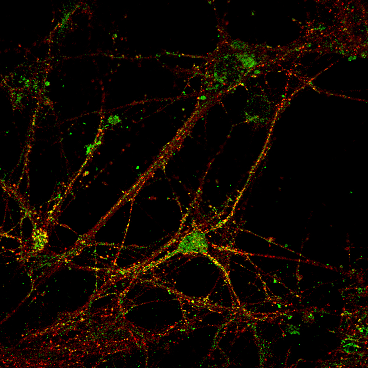
SIM image of hippocampal mouse neuron. NR1 subunit of the NMDA receptor labeled with ATTO 532 and homer labeled with ATTO 647N.
Prof. Dr. Markus Sauer, Department of Biotechnology & Biophysics, Julius-Maximilians-University, Würzburg, Germany.
Prof. Dr. Markus Sauer, Department of Biotechnology & Biophysics, Julius-Maximilians-University, Würzburg, Germany.

Methanol fixed MDA-MB-468 cells; tubulin-antibody staining with mouse IgG anti beta tubulin (primary) and donkey IgG-fab2-fragment anti mouse labeled with ATTO 488 (secondary).
Prof. Dr. Claus Seidel, Molecular Physical Chemistry, Heinrich Heine University, Düsseldorf, Germany.
Prof. Dr. Claus Seidel, Molecular Physical Chemistry, Heinrich Heine University, Düsseldorf, Germany.

dSTORM image of SLAH3 anion channel in HEK cells labeled with ATTO 655.
Prof. Dr. Markus Sauer, Department of Biotechnology & Biophysics, Julius-Maximilians-University, Würzburg, Germany.
Prof. Dr. Markus Sauer, Department of Biotechnology & Biophysics, Julius-Maximilians-University, Würzburg, Germany.

Phase-separated giant-unilamellar-vesicles (equatorial scan) where the liquid-ordered phase is marked with ATTO 488-DPPE (green) and the liquid-disordered phase is labeled with ATTO 647N-DOPE (red).
C. Eggeling et.al., J.Biol.Chem. 291, (2016), 16948
C. Eggeling et.al., J.Biol.Chem. 291, (2016), 16948

Expression of aquaporin 3 in rat colon. The rat colon sections (paraffin-embedded) were stained with anti-aquaporin 3-ATTO 594 anti-body (1:100). Staining (red color) is present in absorptive cells of the colonic epithelium. Hoechst 33342 (blue) is used as counterstain.
Alomone Labs Ltd., Jerusalem, Israel
Alomone Labs Ltd., Jerusalem, Israel

STED: Immunofluorescence of spot-tagged actin-chromobody with ChromoTek‘s Spot-Label® ATTO 594 bivalent (1:1,000). Gated STED image acquired with a Leica TCS SP8 STED 3X microscope with pulsed white light laser excitation at 590 nm and pulsed depletion with a 775 nm laser.
STED image was recorded at the Core Facility Bioimaging at the Biomedical Center, LMU Munich, Germany.
STED image was recorded at the Core Facility Bioimaging at the Biomedical Center, LMU Munich, Germany.

MRC5, Lungfibroblasts: actin labeled with ATTO 430LS phalloidin, alpha tubulin labeled with mouse IgG alpha-tubulin-AK (primary) (green) and ATTO 490LS labeled anti mouse IgG (secondary) (red), nucleous DAPI (blue).
Paul-Ehrlich-Institut (PEI), Darmstadt, Germany.
Paul-Ehrlich-Institut (PEI), Darmstadt, Germany.